Half-Life Extension Strategies for Peptide Therapeutics
Engineers deploy multiple strategies to extend peptide half-life, each with distinct tradeoffs.
Peptide half-life extension has grown into a toolkit of molecular engineering approaches, and each one has its own mechanism, ceiling, and clinical cost. The field has matured past the question of whether to engineer longevity into a peptide and arrived at the harder question of which combination of strategies a given molecule and indication actually demands. This piece maps that toolkit and the tradeoffs that determine how it is deployed.
Why peptides degrade so fast: the three elimination pathways that define the problem
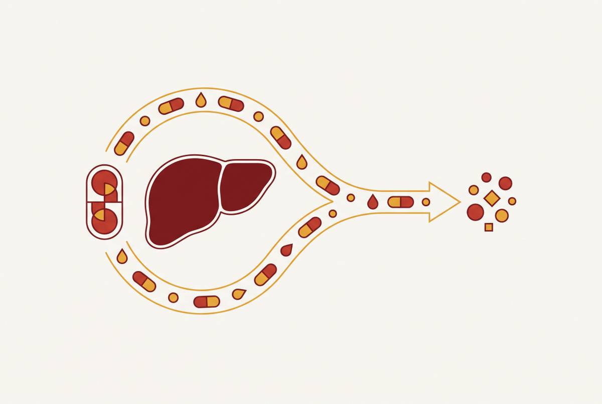
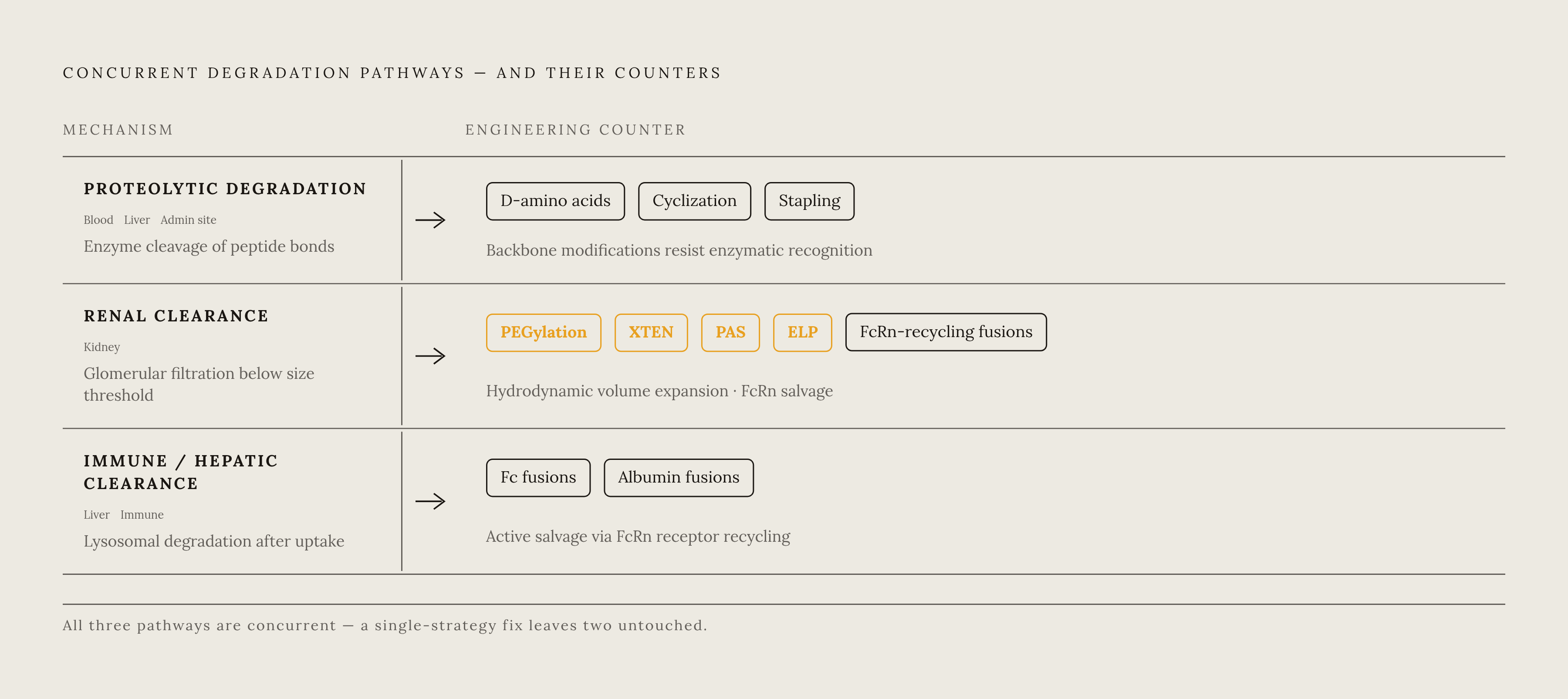
Short peptide half-life is not one defect but the combined result of three distinct elimination mechanisms operating at the same time. The first is proteolytic degradation. Endogenous proteases in blood, liver, and at administration sites cleave peptide bonds and inactivate the therapeutic before it can reach its target. A peptide in plasma is under assault from multiple enzyme families at once, each with its own preferred cleavage geometry.
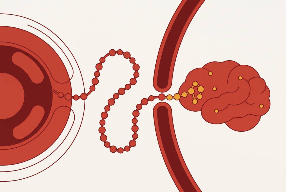
The second mechanism is renal clearance. Peptides with a molecular mass below the glomerular filtration threshold pass freely into urine, and this filtration is driven primarily by small size, independent of any protease activity. A molecule that somehow evaded every protease in the bloodstream would still be eliminated within minutes if its hydrodynamic volume remained below that threshold. Size alone is sufficient grounds for excretion.
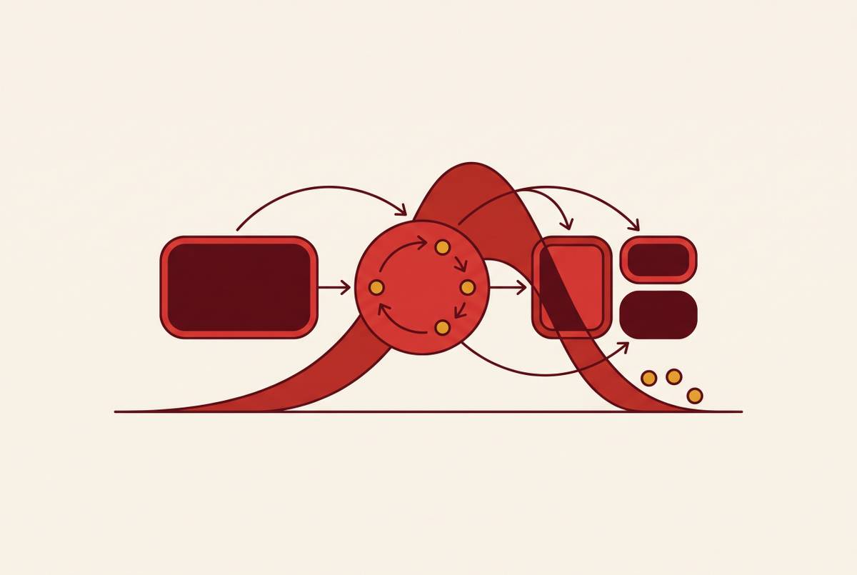
The third mechanism is immune and hepatic clearance. Because all three operate concurrently, a strategy that addresses only one leaves the other two fully intact. That is why the field has converged on multi-layered approaches rather than a single fix.
The scale of the problem becomes concrete in a benchmark molecule. Native GLP-1 carries a half-life of approximately 2 minutes, limited by DPP-4 cleavage at position 2 and rapid renal filtration. Clinical consequences flow directly from the unmodified state: frequent or daily injections, peak-and-trough concentration swings that create windows of subtherapeutic exposure or toxicity, higher doses to compensate, and elevated treatment burden in chronic disease. The engineering mandate is not to nudge half-life upward but to close a gap of several orders of magnitude, and no single mechanism can do that alone.
Structural and chemical modifications that stabilize the peptide backbone itself
The most direct way to block proteolytic recognition is to modify the peptide backbone and amino acid sequence before any carrier or delivery system enters the picture. These intrinsic modifications set a stability floor that every other strategy builds on. D-amino acid substitution illustrates the logic. Not every position tolerates inversion without receptor-binding loss, so you need position-by-position empirical mapping, not a template.
N-methylated amino acids work at the recognition step, but in another way. Non-natural amino acids extend the toolkit further: fluorinated residues resist oxidation, conformationally constrained residues limit the flexibility that enzymes exploit, and side-chain modifications can eliminate specific degradation pathways outright. Beta-amino acids and other non-natural linkages go further still, maintaining biological activity while rendering the backbone unrecognizable to most endogenous proteases.
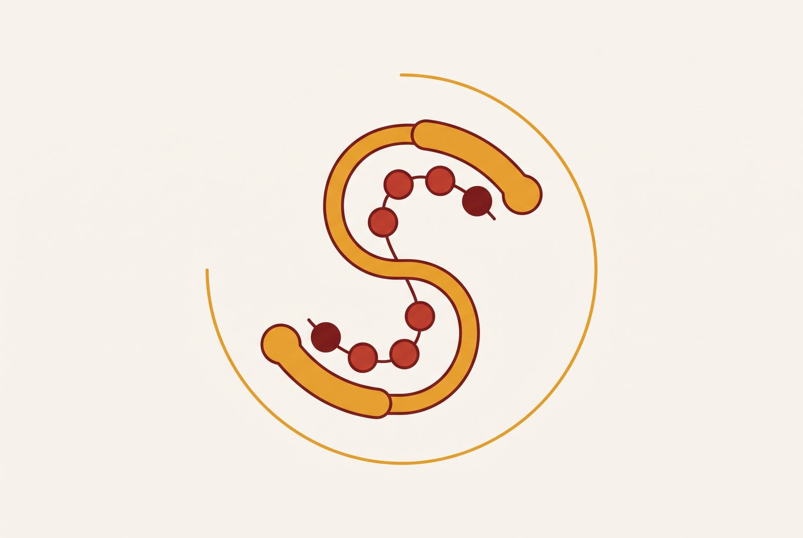
Peptide cyclization addresses exopeptidase entry points directly. Head-to-tail cyclization removes both the N- and C-termini simultaneously, eliminating the two ends that aminopeptidases and carboxypeptidases need to begin. Side-chain-to-side-chain cyclization, whether through disulfide bonds, lactam bridges, or metal coordination, constrains conformation and reduces accessibility of internal cleavage sites. Peptide stapling operates on the same principle through covalent cross-links between side chains, constraining secondary structure, and carries a particular advantage for developing cell-permeable peptides that can access intracellular targets, an indication space most other extension strategies cannot reach.
These chemical modifications act on protease resistance and, to some degree, on renal clearance through conformational compaction. But they do not dramatically increase hydrodynamic volume or recruit recycling receptors. That is what makes them necessary but rarely sufficient for weekly or monthly dosing. The gap they leave is precisely the one the next tier of strategies fills.
Polymer conjugation: how PEGylation and its successors bulk up a peptide to escape renal clearance
Attaching a bulky, hydrophilic polymer to a peptide is the most direct way to push its effective hydrodynamic size above the renal filtration threshold. Polyethylene glycol conjugation increases apparent molecular size far beyond the raw mass of the PEG chain, because PEG binds water and creates a large hydrodynamic shell. That shell blocks glomerular filtration, and it also creates steric hindrance around protease cleavage sites. One conjugation event addresses two of the three elimination pathways at once.
PEGylation offers tunable, predictable half-life extension. Site-specificity of conjugation therefore matters enormously. Random conjugation risks blocking the active site. Site-directed PEGylation, to engineered cysteines or non-natural amino acids, preserves activity while retaining the pharmacokinetic benefit.
Two further tradeoffs matter clinically. Next-generation alternatives such as polysialic acids, hydroxyethyl starch, and other biodegradable polymers are in development to address the immunogenicity and accumulation concerns while retaining the hydrodynamic volume benefit. None has yet achieved the breadth of clinical precedent that PEGylation carries.
The key limitation sits at the level of mechanism. The half-life ceiling it achieves is lower than what you can reach with fusion-based approaches that recruit FcRn or albumin recycling. That distinction, between passive size-based extension and active receptor-mediated salvage, defines the next dividing line in the toolkit.
Fc and albumin fusions: borrowing the body's own recycling machinery for the longest half-lives
Fusing a therapeutic peptide to a carrier protein that the body actively rescues from degradation is the most powerful half-life extension mechanism available. The molecular complexity this creates introduces manufacturing and tissue-penetration costs that smaller modifications avoid, which is why the approach is not universally the right choice. But for ceiling, nothing currently established exceeds it.
The mechanism is the neonatal Fc receptor, FcRn. This receptor binds IgG and albumin in the acidic endosomal compartment at approximately pH 6.0 after cellular uptake, shuttles them back to the cell surface, and releases them at physiological pH 7.4. The salvage pathway continuously rescues Fc-fusion and albumin-fusion proteins from lysosomal degradation, yielding half-lives measured in days to weeks. The clinical proof is dulaglutide, a GLP-1–Fc fusion approved as a weekly injectable, so Fc fusion can convert a minutes-lived peptide into a once-weekly drug.
Human serum albumin fusion offers a parallel route. Direct genetic fusion to HSA transfers that longevity to the therapeutic peptide. An albumin binding domain or AlbudAb domain is fused to the peptide instead, and the resulting construct binds circulating endogenous albumin non-covalently in vivo, hitchhiking on albumin's recycling pathway without requiring albumin to be expressed as part of the fusion protein. This confers extended half-life via FcRn-mediated recycling and avoidance of renal clearance, and it reduces the size of the manufactured product.
Two further fusion approaches round out the class. Carboxy terminal peptide fusion increases hydrodynamic volume through sialylation of the CTP sequence, a distinct mechanism from FcRn recycling that offers moderate extension without the full size of an Fc or albumin fusion. These options offer different size, cost, and recycling-pathway profiles, so you have no single hierarchy.
The costs are real and structural. Protein fusions provide the most dramatic half-life extension by exploiting FcRn recycling. This is the ceiling of systemic half-life extension achievable through any currently established molecular engineering approach, but you get it attached to a large, complex molecule, and that tradeoff motivates the search for middle options.
Unstructured polypeptide tags (XTEN, PAS, ELP) as a biodegradable middle path
Genetically encoded unstructured polypeptide tags achieve half-life extension through hydrodynamic volume expansion without the non-biodegradability of PEG or the full molecular weight of an Fc fusion. Their defining feature is programmability: because they are encoded at the gene level, length can be tuned precisely, making them a middle option between chemical conjugation and full carrier-protein fusion. The mechanism is disordered conformation. Sequences such as XTEN, PAS, and ELP adopt a highly disordered, extended conformation in solution that inflates the apparent hydrodynamic radius far beyond the polypeptide's molecular weight. The glomerulus sees a larger apparent size, so renal clearance drops, and you do not need a structured carrier protein. PAS tags stay low in immunogenicity because they contain only natural amino acids in random-coil conformation, so they present minimal epitopes.
ELP adds a distinctive capability. Elastin-like polypeptide goes beyond simple volume expansion: it shows temperature-responsive phase behavior that has been exploited for depot formation. At physiological temperature, ELP-fused peptides can form subcutaneous depots that release drug slowly. A GLP-1–ELP fusion kept blood glucose down for up to 5 days after one dose in a mouse model, which is about 120-fold longer than native GLP-1. PsTag, a newer variant in this class, has been engineered for high solubility and low immunogenicity.
The advantages over first-generation approaches are specific. Against Fc and albumin fusions, they yield a smaller final molecule for a comparable half-life increment because disordered coil inflates apparent size without adding structured mass. Length can be dialed from tens to hundreds of residues at the gene level, allowing pharmacokineticists to target a specific half-life window without changing the chemistry of the process.
In vivo antibody conjugation and nanoparticle encapsulation: where the frontier is moving
The newest strategies move beyond modifying the peptide molecule itself. They enlist the body's own endogenous proteins or engineered delivery vehicles, so they open tissue-targeted and non-injectable administration that earlier approaches cannot match. In vivo antibody conjugation through the Z33 domain illustrates the shift. The therapeutic peptide is attached to this Z33 warhead, which then self-assembles onto endogenous IgG after subcutaneous injection. Selectivity for IgG subclasses, potential interference with antigen-binding functions of occupied IgG, and scale-up of the electrophilic warhead chemistry are active research areas.
Nanoparticle encapsulation operates on a different logic entirely. The pharmacokinetic profile is determined by formulation rather than molecular engineering of the peptide itself. Surface functionalization of nanoparticles with ligands such as antibody fragments, peptides, or small molecules allows preferential accumulation in target tissues, including the CNS via nose-to-brain routes. And nanoparticle encapsulation is a prerequisite technology for intranasal and oral peptide delivery, because protecting the peptide through the nasal mucosa or gastrointestinal tract requires physical shielding that chemical modification alone cannot provide. Microneedle patches are an emerging self-administration format that combines slow-release depot formation with minimally invasive delivery, so you avoid the needle burden of conventional subcutaneous injection.
What distinguishes this tier is spatial control. Molecular engineering modifies the peptide and accepts systemic distribution as a given. Nanoparticle and in vivo conjugation strategies shape where the peptide goes and how long it persists there, adding a spatial dimension to half-life extension that the molecular toolkit does not possess.
How the tradeoffs stack up: choosing the right strategy for a given peptide and indication
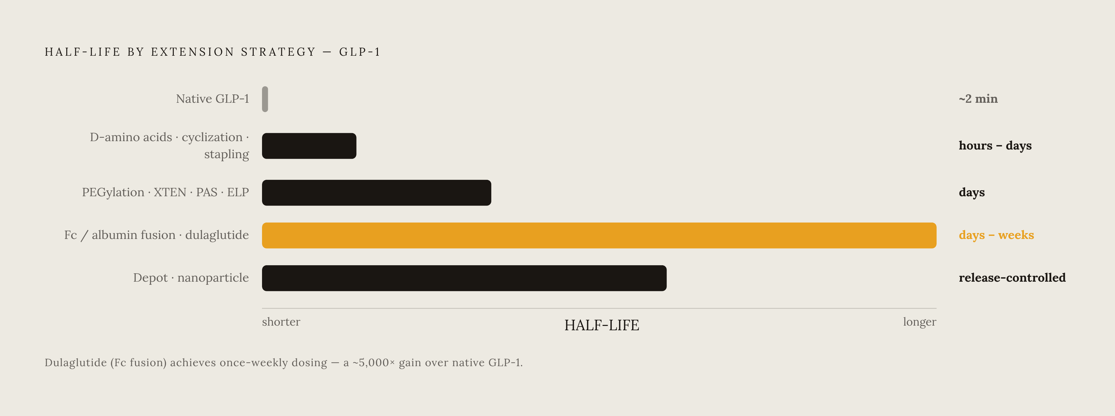
No single half-life extension strategy dominates every situation. The right choice is determined by target half-life, molecule size constraints, route of administration, tissue access requirements, manufacturing feasibility, and the immunogenicity tolerance of the indication. The three core mechanisms have different ceilings, and if you understand those ceilings, you can choose more rationally.
Intrinsic stabilization through D-amino acids, cyclization, and stapling adds hours to days of half-life at low added mass. Hydrodynamic volume expansion through PEGylation, XTEN, PAS, or ELP adds days, with biodegradable options available in the polypeptide tags, moderate size increase, and no active recycling. The ceiling sits below what Fc and albumin achieve. Active recycling through Fc fusion, albumin fusion, or albumin binding domains adds days to weeks at the largest size and manufacturing complexity, with the highest ceiling but with effector function and competition with endogenous IgG as considerations. Physical encapsulation through nanoparticles, liposomes, or depot systems differs in kind: duration is set by formulation release kinetics, not molecular modification, and it is the only class that adds route flexibility and tissue targeting, including enabling non-injectable delivery of otherwise injectable peptides.
Layering is the norm, not the exception. The most successful commercial peptide therapeutics combine multiple approaches: chemical stabilization to block protease cleavage, followed by a carrier or encapsulation strategy to reduce renal clearance and extend circulation. Large Fc and albumin fusions are poorly suited because the blood-brain barrier and blood-CSF barrier sharply limit access for high-molecular-weight constructs. Intrinsic stabilization combined with nanoparticle-enabled nose-to-brain delivery is mechanistically better aligned for reaching brain targets. Chronic metabolic disease pushes the opposite direction: weekly-to-monthly dosing goals favor Fc or albumin fusion, where regulatory precedent exists, or depot-forming ELP and nanoparticle systems. Acute or intermittent indications may justify intrinsic stabilization alone if half-life in the hours-to-days range is acceptable.
Manufacturability functions as a non-negotiable filter. The production pathway must be selected in parallel with the half-life strategy, not after. All carrier strategies introduce non-native structural elements. Fc and albumin are self-proteins with strong tolerogenic precedent. The Z33 in vivo conjugation approach is least characterized immunologically and carries the most open questions.
The patient-access dimension ties the molecular engineering back to its purpose. Half-life extension is not only a pharmacokinetic goal. It is the engineering lever that determines dosing frequency, which in turn determines whether patients actually stay on therapy. Strategies that extend half-life and also enable a less burdensome route, whether oral or intranasal, address both the pharmacokinetic and the compliance problem together. That dual objective, not any single molecular mechanism, is what now separates peptide candidates that reach patients from those that stall in development.
Sources
- Frontiers
- Molecular engineering approaches to half-life extension of therapeutic biomolecules - PubMed
- Peptide and Protein Tags for the Half-Life Extension of Therapeutic Agents: A Review Article - PubMed
- Half-life extension of therapeutics: Applications and mechanisms - PubMed
- Boosting peptide half-life: enabling efficient generation of Fc–peptide conjugates
- Conjugates for use in peptide therapeutics: A systematic review and meta-analysis