CSF Pharmacokinetics and Brain Parenchyma Drug Distribution
CSF presence doesn't guarantee drug reaches the brain tissue where it needs to work.
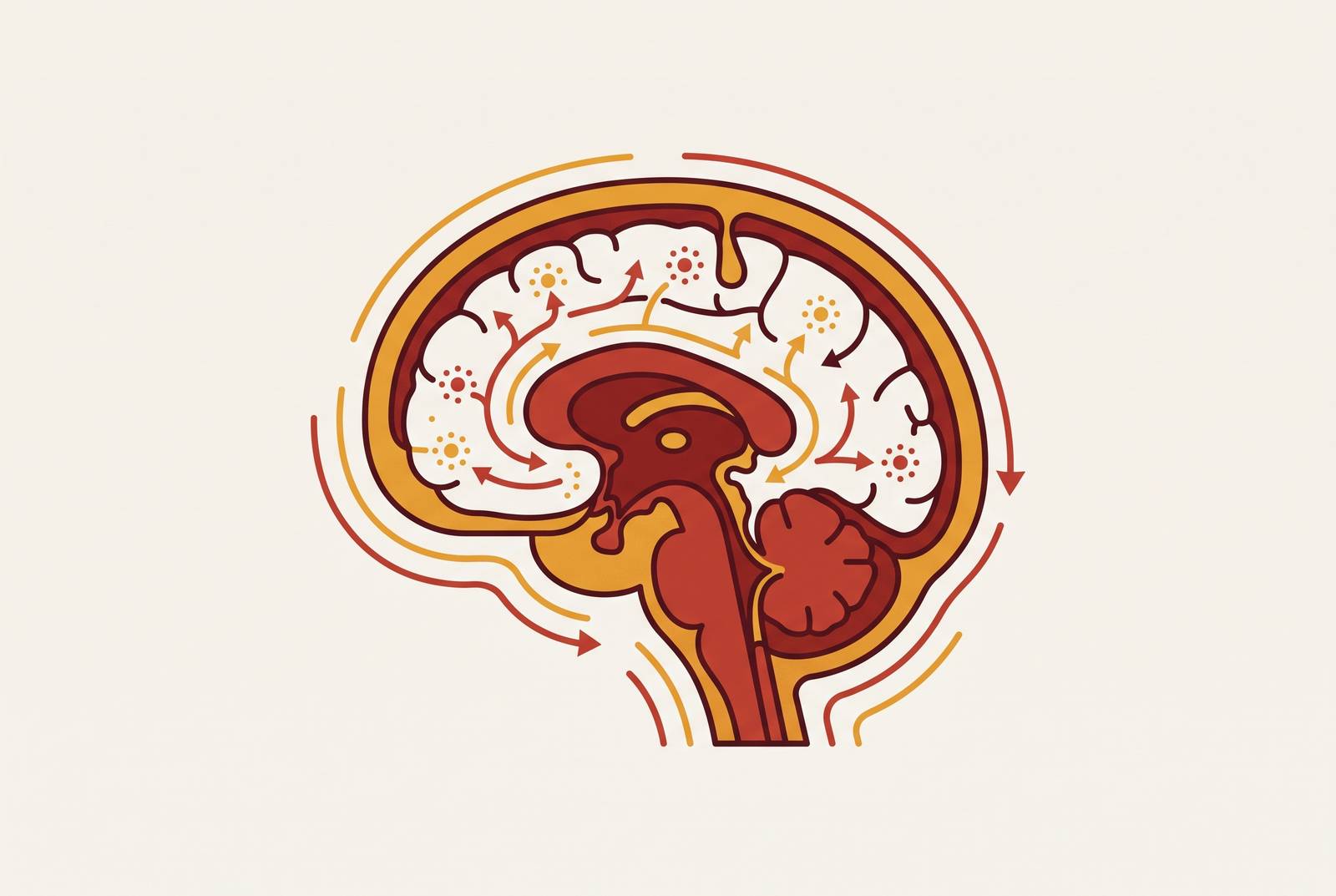
A drug that appears in cerebrospinal fluid has not, by that fact alone, reached the brain. That intuition, common in CNS drug development, treats the brain as a single bucket: cross the blood-brain barrier, find the molecule in spinal fluid, declare victory. The brain actually presents at least three distinct fluid compartments: ventricular CSF, subarachnoid CSF, and brain parenchymal interstitial fluid (ISF), each one governed by its own transport rules. Collapse those three into one, and you get a drug candidate that looks active in early testing but then fails to move the needle in a patient whose disease lives in tissue the CSF sample never represented.
Part of the confusion starts with anatomy that looks similar from a distance but behaves very differently up close. The blood-brain barrier, formed by the microvascular endothelium threading through brain tissue, and the blood-CSF barrier, seated at the choroid plexus, are separate structures with separate transport properties. A drug crossing into CSF and a drug crossing into parenchyma are passing through two different checkpoints, not the same checkpoint sampled at two points in time. Treating CSF concentration as a stand-in for what's happening in the surrounding tissue assumes a connection that the anatomy does not guarantee.
A 2026 review by Engelhard and colleagues makes a point worth sitting with: CSF circulation looks simple on a diagram, but the authors describe it as "amazingly complex," shaped by a long list of interacting factors that determine how fluid and the molecules riding in it actually move through the central nervous system<sup>1</sup><sup>7</sup>. CSF itself is not one well-mixed pool moving uniformly; it has a macrocirculation, bulk flow through the ventricles, cisterns, subarachnoid space, and spinal canal, and it has a microcirculation, localized currents running along ventricular walls, through sulci, and along perivascular channels. These two systems move drug on different timescales and by different physical mechanisms. The sections that follow build out each layer of this architecture in turn: how CSF moves and clears, why that movement undermines the idea of stable drug concentration, how the gap between CSF and parenchyma has been measured, why animal data has made the problem worse rather than better, and what the glymphatic system can and cannot contribute to closing that gap.
How CSF Is Produced, Circulated, and Cleared
CSF is produced, pushed, and drained continuously, driven by a set of overlapping physiological forces: the pulsing of blood vessels, the rhythm of breathing, the coordinated beating of ependymal cilia lining the ventricles, and the pressure gradients that keep the whole intracranial system in balance. Together these forces turn the CSF compartment into a high-turnover environment, one built to move fluid through and out, not to hold anything in place. For a drug sitting in that fluid, this is close to the worst possible storage condition.
Engelhard and colleagues (2026) describe how each heartbeat sends a traveling wave through the choroid plexus and along the CSF pathways, contributing real mechanical force to pushing CSF through the ventricular system and into the subarachnoid space. CSF pressure, flow rate, and absorption are tied tightly to overall brain homeostasis, so this is not a side effect of circulation; it is circulation doing its job. Ependymal cilia add a second, independent driver. Their coordinated beating moves fluid locally along the ventricular walls, separate from vascular pulsatility, so CSF flow has more than one engine behind it. A therapy that tries to slow or trap CSF movement by targeting a single mechanism will run into the other mechanisms still operating.
Imaging has made this picture sharper. Phase-contrast MRI, as Engelhard and colleagues note, has changed what's understood about CNS fluid dynamics by showing that CSF flow shifts with circadian rhythm, with age, with disease state, and with a range of molecular factors. The same patient can present a meaningfully different hydrodynamic environment depending on the time of day a measurement is taken. For drug delivery, the consequence is direct: a molecule placed into CSF, whether by intrathecal injection, intraventricular infusion, or absorption through the olfactory mucosa, enters a system that is already working to transport it away and clear it out. It does not sit and wait for the next sampling point. Bulk flow is an efflux force first, and anything that looks like retention is really a transient snapshot of a molecule in the middle of being removed.
CSF Drug Concentration as a Poor Proxy for Brain Parenchyma Exposure
This is where the central claim of the piece sits: CSF concentration measures CSF, not the tissue the drug is meant to treat. Even when a drug shows up in spinal fluid at a concentration that looks therapeutically reasonable, the parenchyma, the actual brain tissue where the drug needs to act, sits behind diffusion barriers that cause concentration to drop sharply with distance from the CSF surface. CSF sampling is convenient because it is accessible through lumbar puncture or ventricular catheter. Convenience is not the same as relevance to what's happening a few millimeters deeper into tissue.
The scale of this spatial mismatch has been quantified directly. A nine-compartment physiologically based pharmacokinetic model, referred to as the 9-CNS model, was validated against clinical data from glioblastoma patients treated with six different drugs: abemaciclib, ribociclib, pamiparib, olaparib, temuterkib, and ceritinib. The model demonstrated that drug exposure varies substantially and predictably across CSF, parenchyma, tumor rim, bulk tumor, and tumor core, all from the same systemic dose. Different regions of the same brain, in the same patient, routinely see very different concentrations of the same molecule. That is not a modeling artifact; it is what six validated drug cases in real patients showed.
Two separate transport processes drive that variation. Drug movement from CSF into parenchyma happens through passive diffusion and, in some regions, through paravascular convective bulk flow, and these two processes respond to different molecular properties. A molecule's size, charge, and lipophilicity can favor one pathway over the other, so two drugs with identical CSF concentrations can reach very different concentrations once they arrive in tissue. One might argue this is a technical detail best left to pharmacokinetic modelers, but it has a direct clinical consequence: a drug achieving adequate CSF levels can still fail to reach therapeutic concentrations in deep parenchyma, and a drug dismissed as inadequate based on CSF sampling might be performing just fine in the tissue that matters. Standard CSF biosampling cannot catch either error. It reports on the compartment it was drawn from and nothing further in.
The species translation gap that CSF sampling has hidden
The problem with CSF as a proxy does not stay inside a single patient or species. It compounds across the preclinical-to-clinical pipeline, because CSF-to-plasma drug ratios differ systematically between rodents and humans. A concentration ratio that looks favorable in a mouse does not translate cleanly to a human. Rodent CSF data carries its own layer of unreliability stacked on top of the CSF-versus-parenchyma gap already described. Two sources of error compound each other.
Engelhard and colleagues (2026) treat this translation question as one of the central reasons for reviewing CSF circulation variability in the first place, pointing out that comparisons between laboratory animals and humans are necessary precisely because CSF dynamics do not operate the same way across species. Disease adds a further complication on top of species difference. Pathological states alter CSF dynamics and barrier integrity in ways that a healthy animal model simply does not reproduce, so translation failures are not purely a matter of rodent versus human physiology. They also reflect the gap between a disease-free animal and a human patient whose condition has already reshaped the fluid system being studied.
None of this reflects poorly on the researchers who built CNS drug pipelines around CSF sampling over the past several decades. CSF was the measurement available, accessible, and clinically standard. But the compounding of species bias and disease-state mismatch helps explain a specific, recurring pattern: CNS drug candidates that perform well against preclinical CSF benchmarks and then underperform once they reach human trials. That pattern is why mechanistic, compartment-explicit modeling, of the kind built into the 9-CNS PBPK framework, matters now in a way it could not have a generation ago. It does not replace CSF sampling so much as correct for the systematic bias that sampling alone cannot reveal.
The glymphatic system as the bridge between CSF and parenchymal ISF, and its limits as a drug delivery pathway
If CSF and parenchyma are separated by diffusion barriers and distance, something still has to move fluid, and whatever rides in it, from one compartment to the other under normal physiology. That bridge is the glymphatic system, a pathway that exchanges fluid between CSF and parenchymal ISF through paravascular convective flow. The flow is driven by arterial pulsatility, by the respiratory cycle, and by pressure gradients in the CSF itself. It is the clearest mechanistic link available between the two compartments this article has spent several sections separating.
The pathway was defined formally in a patent issued to Nedergaard and colleagues in 2018 (US Patent 9,901,650), which describes the glymphatic system as a brain-wide paravascular route for waste clearance, linking CSF and ISF exchange in ways that can be measured through imaging. That patent gave the field both a name for the mechanism and a method for tracking it. But naming a pathway is not the same as controlling it for drug delivery purposes. Paravascular bulk flow depends on arterial pulsations that expand vessel walls and push fluid forward in a peristaltic rhythm, and that rhythm shifts with sleep state, with arousal, and with disease, the same variables that already complicate CSF bulk flow described earlier in this piece. A drug that enters CSF and hopes to ride the glymphatic system into parenchyma is depending on a biological process with its own regulators, awake, asleep, healthy, or diseased, rather than on a fixed, engineerable channel.
Engelhard and colleagues (2026) reinforce this by describing CSF microcirculation, including the perivascular pathways the glymphatic system depends on, as distinct from macrocirculation, local and variable. A molecule can reach CSF-exposed surfaces of the brain and spinal cord, and through glymphatic-associated movement it can travel some distance into parenchyma. But the ependymal cell layer associated with the choroid plexus remains a limiting barrier to drug action within the tissue, even after intrathecal administration places a drug directly into CSF. Glymphatic access narrows the gap between CSF and parenchyma. It does not dissolve it, and the next section turns to what closing that remaining gap actually demands of a drug and its formulation.
Engineering requirements for reaching deep brain parenchyma
Diagnosing the gap between CSF and parenchyma is one thing. Closing it is another, and closing it requires treating the problem as one of molecular and formulation design rather than one solved by picking the right injection site. Reaching meaningful concentrations in deep brain parenchyma means managing at least four properties simultaneously: passive permeability across the blood-brain barrier, the activity of efflux transporters working against the drug at that barrier, how much of the drug is bound to protein rather than free to act, and the diffusion distance separating the target tissue from the nearest CSF surface. No single route of administration optimizes all four at once.
Work by Li and colleagues shows that CNS drug distribution is driven by the plasma concentration-time profile, shaped jointly by the drug's own properties and by the pathophysiology of the CNS being treated. Neither dose nor route alone decides what reaches parenchyma. Molecular properties interact with the anatomical system at every step along the way. The 9-CNS PBPK model builds in both passive permeability and transporter-mediated active efflux or uptake clearance at the blood-brain barrier, predicted from laboratory data through in vitro-in vivo extrapolation. Efflux transporters sitting at the BBB endothelium actively work against molecules that have already entered passively, so you have to design for a drug's interaction with those transporters in advance, not discover it as a disappointing result after a trial has run.
For drugs given systemically, through an intravenous line or an oral dose, the plasma concentration-time profile is the main driver of whatever exposure the CNS eventually sees. But how does this affect the original promise of a drug with excellent CNS-penetrating properties on paper? Even such a molecule still has to achieve and hold adequate plasma levels, with the exposure window at the parenchyma set by where plasma pharmacokinetics and CNS transport rates intersect. Intrathecal and intraventricular routes sidestep that peripheral pharmacokinetic problem by placing the drug directly into CSF, but they trade it for the variability already described in CSF circulation itself, affected by cardiac cycle, age, posture, and disease. Drug distribution from CSF into parenchyma after direct CNS administration is, in its own right, inconsistent and hard to predict without characterizing an individual patient's flow pattern. Bypassing one obstacle does not remove the need to engineer around the next one.
The Nose-to-Brain Route and the Geometry of the CNS Delivery Problem
Intranasal delivery approaches the problem from a different angle. Rather than placing a drug into systemic circulation or directly into CSF and hoping it eventually reaches parenchyma, the olfactory and trigeminal nerve pathways deposit drug directly into CNS tissue close to cortical and subcortical targets. That geometry partially decouples delivery from the CSF bulk-flow efflux problem described earlier in this piece, but only under two conditions: the drug has to survive nasal transit long enough to be absorbed, and it has to be able to reach targets that extend beyond the olfactory bulb itself.
Drug absorbed by the olfactory epithelium travels along olfactory nerve fibers to the olfactory bulb, and from there it can distribute to the cerebral cortex, the hippocampus, and other brain regions through CSF, through interstitial fluid, and through transsynaptic pathways along neural connections. That trajectory bypasses the blood-brain barrier altogether and enters parenchyma at the olfactory bulb rather than diffusing inward from a CSF surface the way an intrathecally delivered drug would. The trigeminal nerve pathway extends that reach further, giving access to deeper CNS structures and complementing what olfactory transport alone can achieve.
None of this comes free of its own kinetic constraints. The primary obstacle facing the nasal route is mucociliary clearance, the same kind of constant physical movement that limits how long anything sits in the nasal cavity before being swept away. That limits how long a drug has to be absorbed before it is cleared. Dosing volume compounds the constraint: the maximum volume deliverable in humans is 1 mL per nostril, with an ideal dosing volume closer to 0.1 to 0.15 mL per nostril. That is a narrow window to work within, and it means the amount of drug that can be delivered in a single administration is tightly capped. Formulation efficiency, how much active drug can be packed into a small volume and how well it survives the trip across the nasal mucosa, is not an optional refinement.
Sources
- TYPE Review PUBLISHED 12 May 2026 DOI 10.3389/fddev.2026.1735474
- Mechanistic Modeling of Spatial Heterogeneity of Drug Penetration and Exposure in the Human Central Nervous System and Brain Tumors - PMC
- CSF, blood-brain barrier, and brain drug delivery - PubMed
- Methods for evaluating brain-wide paravascular pathway for waste clearance function and methods for treating neurodegenerative disorders based thereon
- Intranasal Drug Delivery Technology in the Treatment of Central Nervous System Diseases: Challenges, Advances, and Future Research Directions
- The Nasal–Brain Drug Delivery Route: Mechanisms and Applications to Central Nervous System Diseases