GLP-1 Receptor Distribution in the Human Brain
Brain GLP-1 receptors cluster in cortex, not cerebellum, unlike rodents.

GLP-1 has a reputation problem, in the sense that most people file it under "gut hormone" or "diabetes drug" and stop there. That framing misses something anatomically important: the brain runs its own GLP-1 system, built on its own wiring, and the geography of that system, where the receptors sit and where they do not, is what actually determines what GLP-1 agonism can do to cognition, appetite, mood, and eventually how it should be delivered.
The human brain's own GLP-1 system, separate from the gut
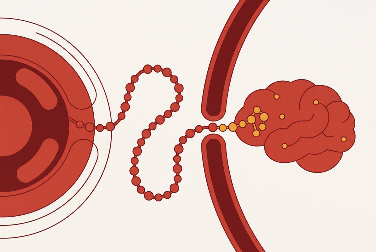
The gut version of the story is familiar. Nutrients hit the intestine, enteroendocrine cells release GLP-1, insulin gets a nudge, and the stomach empties more slowly. But the brain does not wait for that signal to arrive from below. It manufactures its own supply, using the same gene that codes for the gut hormone, expressed instead in a distinct population of neurons in the brainstem's nucleus of the solitary tract, or NTS.
The answer is that it mostly does not. Brain-derived GLP-1, not the circulating gut hormone, is the physiological agonist acting on the brain's own GLP-1 receptors, because peripheral GLP-1 is degraded too fast and crosses into the brain in only limited amounts. The PPG neurons that make it send axons outward toward the limbic system and other regions, laying down what amounts to an internal signaling network that runs on different logic than the endocrine system does. Endocrine signaling floods the bloodstream and waits for receptors to catch what they can. Neuronal signaling is targeted, projecting to specific destinations along specific fibers.
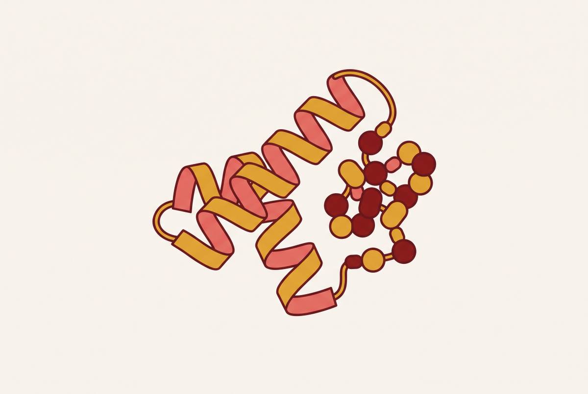
At the receptor level, the mechanism is fairly standard for this class of hormone. The primary intracellular signaling pathway runs through Gs-mediated activation of adenylyl cyclase, which drives rapid cAMP accumulation and activates protein kinase A (PKA). That cascade does more than manage blood sugar or appetite. It appears to carry anti-inflammatory effects and improvements in mitochondrial function, a pleiotropic reach that has made GLP-1 receptor biology interesting well beyond metabolic disease.
None of this means peripheral signals are irrelevant to the brain. They do reach it, just through narrow doors: the circulation, entering at circumventricular organs where the blood-brain barrier is naturally leaky, and the vagus nerve, both of which concentrate their input in the NTS and the neighboring area postrema. Keep that detail in mind. It becomes central later, when the discussion turns to why injectable drugs produce nausea before they produce much else.
The human brain GLP-1 receptor map versus the animal data
If brain GLP-1 operates on its own logic, the next question is obvious: where in the brain does that logic actually play out? The anatomy carries the argument here, because the human receptor map looks nothing like what decades of rodent research would have predicted.
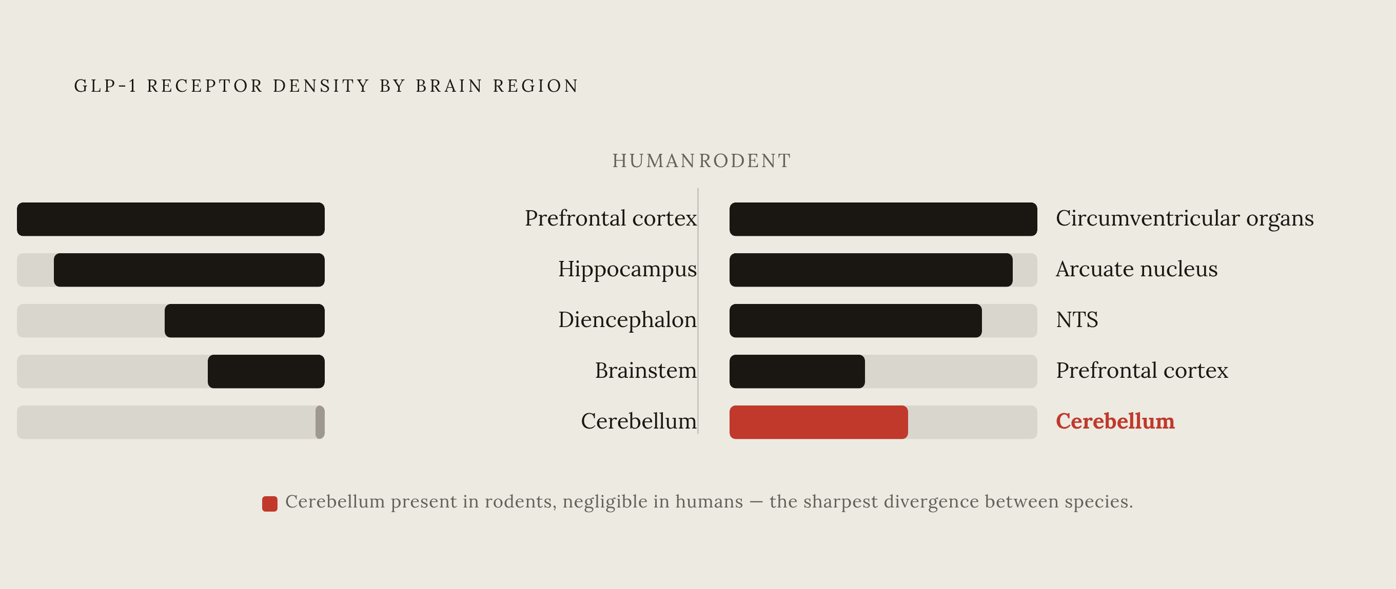
Key human findings come from a whole-brain mapping study across 30 post-mortem human brains, cited in Licinio and Wong, Brain Med. GLP-1 expression shows up across most of the cortex, with the heaviest concentration in the frontal, prefrontal, and parietal regions, and GLP-1 receptor expression peaks specifically in the frontal cortex. The hippocampus, seat of memory formation, carries a substantial receptor presence of its own. Further down, the diencephalon and brainstem also express GLP-1, extending the map into structures involved in basic regulatory functions.
Then there's the cerebellum, which shows essentially nothing. Receptor expression there is negligible enough to count as an absence. For a region long associated mostly with motor coordination, that might sound unremarkable. But it becomes the detail that separates the human map from every rodent map that came before it.
Because in rodents, the pattern runs almost in reverse. In rodents, by contrast, Farkas et al., Brain Struct Funct, 2021, found the highest GLP-1 receptor density in the circumventricular organs, the arcuate nucleus, and the NTS, with GLP-1 receptors also present in the cerebellum, a profile meaningfully different from the human cortical-dominant pattern. Where humans show a cortex-heavy, cerebellum-absent map, rodents show close to the opposite. GLP-1 receptors were found not just on cell bodies and dendrites, where signals are typically received, but on axon terminals and varicosities, hinting at a presynaptic role in modulating neurotransmitter release. Whether that presynaptic function carries over into human neurons is not yet established, and the mapping data available so far cannot answer it.
What can be said is more consequential for anyone designing a therapeutic around this biology. Rodent studies have been the backbone of preclinical GLP-1 research on brain effects for years, and if the receptor geography genuinely diverges by region between species, then rodent data may be systematically pointing at the wrong human targets. A treatment that looks promising in a mouse hypothalamus is not obviously informative about what happens in a human frontal cortex. Score one for anatomy over extrapolation.
Cognition, appetite, and mood effects predicted by the receptor map
Once the map is in hand, the next step is asking what it explains. Receptor density in the frontal cortex and hippocampus connects directly to clinical experience. It is the physical basis underneath cognitive, appetite, and mood effects that have already been observed in patients taking these drugs.
Start with the frontal and prefrontal cortex. These regions handle executive function: impulse control, planning, the moment-to-moment weighing of whether to act on a craving. Dense receptor presence there gives a plausible anatomical account for something clinicians and patients alike have reported, a shift in food-seeking behavior and in food preference that goes beyond simple caloric restriction. It suggests the drugs are not just slowing digestion. They may be altering the decision-making circuitry that decides whether to reach for the food in the first place.
Move to the hippocampus, which specializes in episodic memory and in tying experience to context. Its significant receptor presence lines up with reports of altered memory and cognitive performance under GLP-1 agonism, and it is part of why neuroprotection has become a serious hypothesis in dementia research rather than a speculative aside. Whether that hypothesis holds up under later scrutiny is a separate question, addressed in the next section, but the anatomical plausibility is there.
Then there's the brainstem, home to the NTS and area postrema, the same structures that catch peripheral GLP-1 signals arriving through the bloodstream and vagus nerve. These brainstem regions are the primary entry point for peripheral GLP-1 signals and are directly involved in nausea, satiety signaling, and visceral sensation, the same circuitry that drives the gastrointestinal side effects of current injectable GLP-1 agonists, as Beutler notes in JCI, 2026, that aversive GI side effects of GLP-1 receptor agonists are CNS-mediated. That single fact reframes a widespread assumption. Appetite suppression under GLP-1 receptor agonists has long been credited to peripheral satiety signaling, the stomach telling the brain it's full. Beutler's review in JCI, 2026, makes the case that GLP-1 receptor agonist efficacy in obesity treatment depends critically on reduced energy intake driven from the CNS side of the loop, not the peripheral satiety signaling long credited for the effect.
There is also a reward component: mesolimbic and mesocortical circuits appear to shape how strongly food rewards register. Mesolimbic and mesocortical GLP-1 receptor circuits appear to modulate reward processing, offering a mechanistic account for the blunted response to highly palatable food that many patients describe informally as reduced "food noise". It is a small phrase, but it points at something the receptor map makes literal: quieting a signal that runs through mesocortical and mesolimbic structures, not merely trimming portion sizes.
Put these three regions side by side and a pattern appears. Cortex governs the decision to eat, hippocampus governs the memory and context around eating, brainstem governs the visceral consequence of having eaten. That three-part division doesn't just describe appetite. It offers a working framework for thinking about mood and addiction circuitry too, since the same reward pathways implicated in food reward overlap substantially with circuits studied in substance use.
The age-related receptor decline and the case for earlier intervention
Anatomy does not sit still over a lifetime, and this is where the map stops being a snapshot and starts behaving like a clock. GLP-1 expression in the human brain declines with age, and it does so in a pattern specific enough to suggest a genuine window of opportunity for CNS-targeted GLP-1 therapy, one that narrows as the years go by.
The same post-mortem mapping work cited earlier found that GLP-1 expression drops off across most brain regions after the fifth decade of life. By age 60, the decline has gone further still: GLP-1 disappears from every cortical region except the prefrontal cortex, though it hangs on in sub-cortical structures. That is a striking asymmetry. The cortical regions carrying the most functionally interesting receptor density, the ones tied to cognition and appetite regulation in the previous section, are exactly the regions losing expression fastest.
That raises an important question for anyone thinking about treatment timing. If cortical GLP-1 signaling is what underlies cognitive and metabolic benefit, and if that signaling is eroding by the sixth decade of life, then an intervention aimed at those same cortical targets presumably works best while the receptors are still there to reach. The implication splits in two directions. For metabolic disease, earlier treatment could preserve more of the CNS-mediated benefit before cortical expression fades. For neurodegenerative disease, the timing question is an anatomical constraint as well as a matter of clinical judgment: treating a hippocampus and frontal cortex that have already lost most of their receptor density is a fundamentally different proposition than treating one that has not.
That distinction matters for how later-stage dementia trials involving GLP-1 compounds ought to be read. A therapy targeting receptors that have already thinned out by the time symptoms are severe enough to enroll a patient is not being given a fair test of what it might do earlier in the decline. The anatomy suggests the disappointing result and the unproven hypothesis are not necessarily the same thing.
Why injectable GLP-1 agonists reach the brain poorly
The delivery method behind almost every GLP-1 drug on the market today, subcutaneous injection, was engineered around the peripheral incretin effect: insulin, glucagon, gastric emptying. It was not built with the cortical and hippocampal receptor fields in mind, because those fields were not yet mapped when the injection route became standard. The engineering itself is not at fault; the mismatch is between when the drug delivery decision was made and when the anatomical picture became clear.
The physical obstacle is the blood-brain barrier. GLP-1 peptides are large molecules, and large molecules cross the barrier badly. Researchers at Imperial College London have flagged the resulting low CNS drug concentrations as a real limitation for applications in weight management, cognition, and addiction treatment. Layered on top of that is a practical one: Patel and Niazi, writing in Pharmaceutics in 2025, describe the subcutaneous route itself as invasive in a way that drags down adherence and invites peripheral side effects.
The adherence numbers back that up with real consequence. One cohort study of patients starting GLP-1 receptor agonists found close to half of those with type 2 diabetes, and closer to two-thirds of those without it, had stopped treatment within a year. A registry study out of Karolinska and Copenhagen tracking new users found cumulative discontinuation nearing a quarter at the one-year mark and topping a third by year three. A 2025 dataset from ISPOR, drawing on Truveta data, isolated the reasons behind documented discontinuations and found side effects responsible for 28.2% of all cases, the single largest driver, with cost trailing behind as a secondary factor.
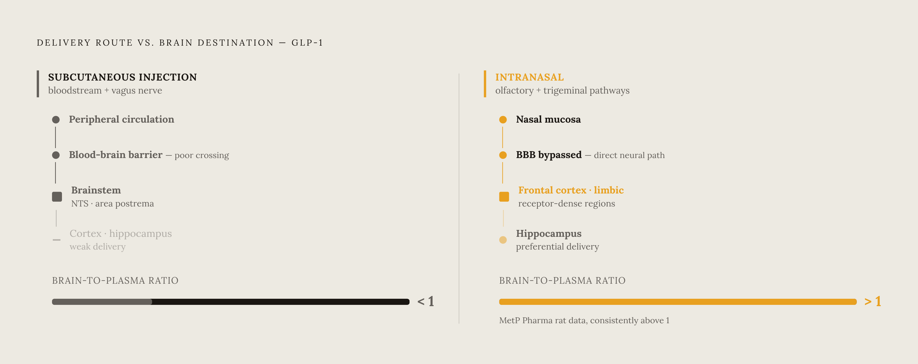
Here the receptor map earns its keep as an explanatory tool rather than just a descriptive one. Nausea, the side effect most often cited by patients who quit, is CNS-mediated, generated through the NTS and area postrema, the same brainstem regions that serve as the entry point for peripheral signals arriving via the bloodstream and vagus nerve. Injection delivers a strong, direct hit to that brainstem gateway while delivering comparatively little to the cortical and hippocampal fields where the map places the receptors tied to cognitive and appetitive benefit. The geography makes the mismatch legible in a way that symptom reporting alone never could.
One might object that injection clearly produces some central effect or these drugs would not suppress appetite at all, and that objection is fair as far as it goes. But it is blunt engagement, concentrated at the brainstem's entry points rather than spread across the frontal and hippocampal fields the map identifies as functionally important. The relevant question is whether the signal that reaches the brain through injection lands in the right regions at doses strong enough to matter.
The nose-to-brain route as a path designed around the receptor map
If the injection route over-delivers to the brainstem and under-delivers to the cortex, the anatomically sensible fix is a delivery path that runs the opposite direction, entering through channels that lead toward the cortex and limbic system rather than toward the brainstem alone. Intranasal delivery, using the olfactory and trigeminal nerve pathways, does roughly that. It sidesteps the blood-brain barrier altogether and opens a route into the exact cortical and limbic territory the human receptor map marks as densest.
The olfactory pathway runs from the olfactory epithelium, which sits in close anatomical range of the olfactory bulb, with projections extending from there toward the frontal cortex and limbic structures, the same regions carrying the heaviest receptor load in the human map. The trigeminal pathway offers a second route and some striking evidence of speed. In one study, a GLP-2 derivative turned up in the trigeminal principal sensory nucleus of the pons just three minutes after intranasal dosing, a fast enough transit time to demonstrate how quickly intra-axonal transport can move material along this route.
Formulation engineering makes that transport possible. Particle size gets treated as a critical variable in this literature, though a 2026 review in Nanoscale is careful to note that small particle size by itself does not guarantee better neuronal transport. Formulation design, not particle size alone, determines whether the payload actually reaches its target. Researchers at Columbia University have developed nanoparticle platforms drawing on more than 30 years of patented delivery work.
There is at least one concrete data point suggesting the approach can work as intended. MetP Pharma reported that its proprietary intranasal delivery technology produced brain-to-plasma ratios consistently above 1 in rats, evidence of preferential delivery to the CNS alongside low systemic exposure, per coverage in Manufacturing Chemist. A ratio above 1 means more drug ends up in the brain than in the blood, which is close to the opposite of what happens with an injected peptide struggling to cross the barrier at all.
As of September 2026, no GLP-1 nasal drug has cleared regulatory approval anywhere. Landmark Medicines Ltd. announced plans to advance its nasal delivery platform toward a human volunteer study, with semaglutide among the candidate molecules under evaluation, citing patient demand for alternatives to injection as the rationale. The pipeline is early. The rationale behind it reflects the anatomical mismatch the receptor map exposes, not a convenience argument dressed up as science. It aims a delivery route at the olfactory and cortical entry points because the human data places the receptors that matter for cognition and appetite regulation there, unlike a route that reaches the brainstem well and the cortex barely.
Sources
- The Multifaceted Nature of GLP-1: Molecular Mechanisms and Signaling Pathways in Metabolic and Neurodegenerative Diseases - PMC
- The brain geography of GLP-1: An atlas for a new era - PMC
- Emerging Frontiers in GLP-1 Therapeutics: A Comprehensive Evidence Base (2025) - PMC
- JCI - GLP-1 physiology and pharmacology along the gut-brain axis
- Brain-Derived GLP-1—Understanding the Physiological Function and Anti-obesity Potential of Preproglucagon Neurons | Endocrinology | Oxford Academic
- Distribution and ultrastructural localization of the glucagon-like peptide-1 receptor (GLP-1R) in the rat brain
- Glucagon-like Peptide-1 Receptor in the Human Hypothalamus Is Associated with Body Mass Index and Colocalizes with the Anorexigenic Neuropeptide Nucleobindin-2/Nesfatin-1
- MetP Pharma unveils brain-targeted intranasal GLP-1 delivery technology