Intranasal GLP-1 Delivery for Addiction and Reward Circuit Modulation
Intranasal delivery bypasses the blood-brain barrier to reach addiction's core neural circuits.

Addiction is often described as a disorder of choice or willpower, but its biology runs through a specific, mappable circuit in the brain, and GLP-1 receptors sit directly inside that circuit. This piece traces that anatomy, shows why current injectable GLP-1 drugs reach those structures only poorly, and lays out why intranasal delivery is the mechanistically coherent route to the ventral tegmental area and nucleus accumbens rather than a convenience upgrade on the current standard.
Where GLP-1 receptors sit in the reward circuit
The mesolimbic dopamine circuit runs through key structures that, together, encode reward salience and drive compulsive seeking behavior: the ventral tegmental area (VTA), the nucleus accumbens core and shell (NAc), and the hypothalamus, the circuitry that assigns salience to a drink, a drug, or a plate of food and drives the compulsive seeking behavior that defines addiction. GLP-1 is not a visitor to this system. Neurons that produce GLP-1 in the nucleus tractus solitarius send monosynaptic projections straight into the VTA and into both the core and shell of the NAc, and these projections fire in response to satiety signals tied to food intake. That wiring gives GLP-1 a direct anatomical foothold in reward processing, not an indirect or hormonal one.
The effect is not confined to a single neurotransmitter system, either. Experimental work shows GLP-1 receptor activation shapes dopaminergic, glutamatergic, and GABAergic signaling across mesolimbic and corticolimbic circuits simultaneously. In the VTA specifically, GLP-1 signaling blunts the phasic dopamine bursts that fire in response to cues predicting food, and in the NAc it dampens the dopamine release normally triggered by drugs of abuse. The reach extends further still: a habenulo–interpeduncular pathway from the NTS, regulated by GLP-1 signaling, is critically involved in nicotine avoidance and aversion, extending the receptor map beyond food and alcohol.
This is a circuit-level fact about precise anatomy, not a loose correlation between a metabolic hormone and appetite. The VTA, the NAc, and the habenulo-interpeduncular pathway are precise coordinates, not metaphors for "the brain's reward system" in the abstract. That precision raises an obvious question for anyone evaluating a GLP-1 drug as an addiction treatment: if the target sits at these exact coordinates, how reliably does the drug now in clinical use actually get there?
What injected GLP-1s do to the reward circuit
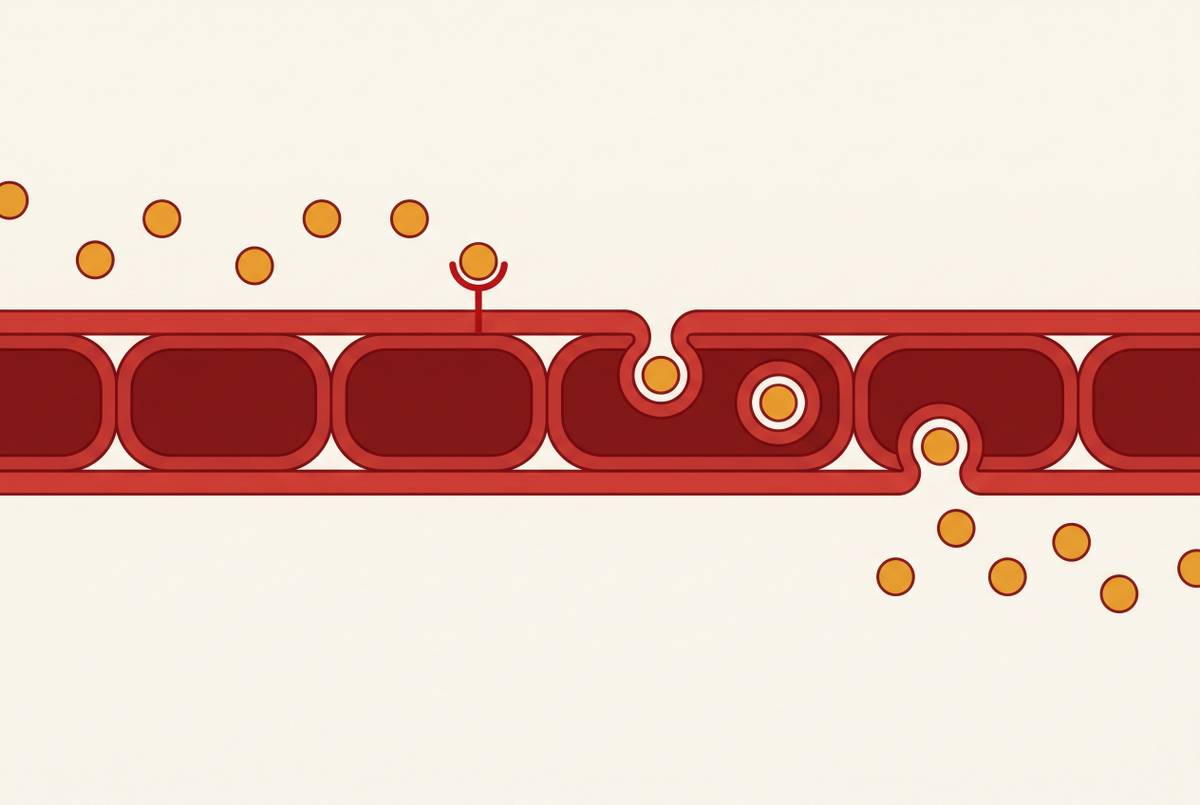
Subcutaneous and oral GLP-1 receptor agonists do what they are designed to do in the bloodstream. They generate high plasma concentrations, and some of that circulating drug eventually crosses into the brain, but only after it clears the restrictive blood-brain barrier, a membrane system built precisely to keep most circulating molecules out. Preclinical studies tracking subcutaneous and oral dosing consistently find brain-to-plasma ratios well below 1, so the concentration reaching brain tissue is a small fraction of what is circulating everywhere else in the body. High plasma exposure does not equal high brain exposure, and the gap between them is a structural feature of how subcutaneous delivery works; it is not an incidental side effect of any one molecule.
That gap has a visible downstream cost. Because so much of the dose stays in the periphery, drug saturates the gut where it was never meant to pool, and that saturation drives the gastrointestinal side-effect profile, nausea and diarrhea, documented so reliably in these drugs' clinical experience. One might argue the behavioral evidence settles the question anyway, since injectable GLP-1s have shown real effects on reward-driven behavior: reduced alcohol craving, attenuated compulsive food seeking, and measurable drops in purchases of processed foods, sugar-sweetened beverages, refined grains, and beef. But that argument cuts the other way as much as it supports the drug. Those effects are arriving despite a delivery route that gets only a sliver of the dose to the VTA and NAc, which suggests the full strength of the signal is being muted before it ever reaches its target.
The clearest demonstration of what more brain exposure can do comes from outside the addiction literature entirely, in neurodegeneration research. The ELAD trial, run by Imperial College London in non-diabetic patients with mild to moderate Alzheimer's disease, tested 52 weeks of daily injected liraglutide in a phase 2 study; it found nearly half less brain volume loss and measurably slower cognitive decline, and the results were published in Nature in December 2025. Liraglutide's pharmacokinetics give it somewhat closer CNS proximity than oral semaglutide achieves, and that difference in exposure, not a difference in which molecule was used, is the most direct explanation for why one program showed a strong CNS signal and another has struggled to. Delivery route answers how much of any given molecule actually arrives where the VTA and NAc can use it, not which GLP-1 molecule is best.
Nose-to-brain route to the VTA and NAc
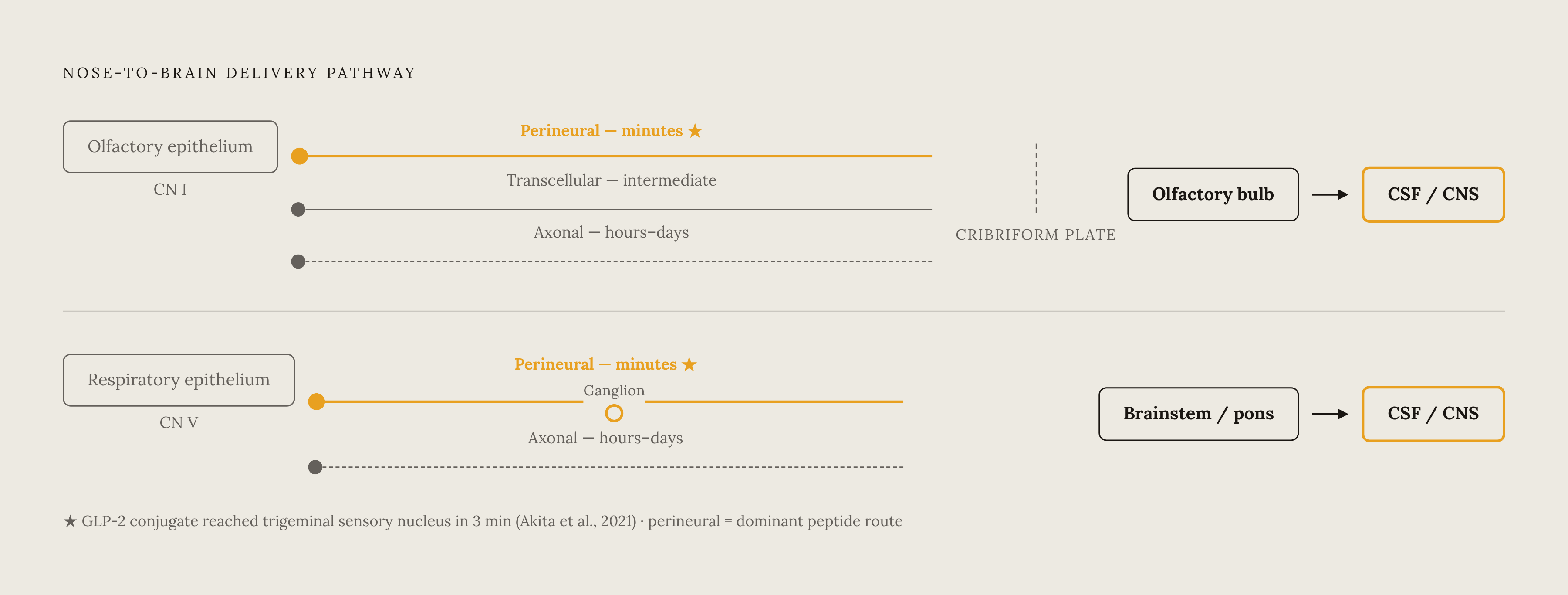
If the obstacle is the blood-brain barrier, the logical move is to find a path that avoids it rather than one that tries to cross it more efficiently. The olfactory and trigeminal nerves offer exactly that kind of path, a direct anatomical connection running from the nasal mucosa into the central nervous system that sidesteps the barrier altogether, carrying molecules into brain tissue within minutes of dosing. Two cranial nerves do the work. The olfactory nerve, cranial nerve I, runs from the olfactory epithelium through the cribriform plate into the olfactory bulb, while the trigeminal nerve, cranial nerve V, innervates the respiratory epithelium and connects to the brainstem by way of the trigeminal ganglion.
Three mechanisms move molecules along these nerves. Intracellular axonal transport is the slowest, taking hours to days; transcellular transport through supporting cells is another route; and extracellular perineural transport is the fastest, acting on a timescale of minutes and dominating for most peptide-sized molecules. The perineural pathway connects directly into the cerebrospinal fluid of the subarachnoid space, so peptides given intranasally can turn up in CSF within minutes of dosing. The FDA's approval of intranasal esketamine for treatment-resistant major depressive disorder established, in a real clinical setting, that the nose-to-brain route can carry a CNS-active drug to therapeutic concentrations in humans.
For GLP-1 peptides specifically, the relevant evidence comes from a close relative in the same peptide family. A GLP-2 derivative conjugated to a cell-penetrating peptide reached the trigeminal sensory nucleus within 3 minutes of intranasal dosing in mice, in work published by Akita and colleagues in the Journal of Controlled Release in 2021. That speed matters because it confirms GLP-family peptides can use the perineural pathway on a timescale fast enough to be preclinically meaningful, not just theoretically possible. Together, the olfactory route's reach into the olfactory bulb, hippocampus, and cortex and the trigeminal route's reach into the brainstem and pons cover both the upstream and downstream anatomy of the mesolimbic circuit mapped in the first section. The nose is a second, anatomically legitimate door into the same circuit the injection struggles to reach.
Why intranasal GLP-1 delivery is harder than it sounds
Anatomical availability is not the same as pharmaceutical accessibility. The nose-to-brain pathway exists in every human nasal cavity, but GLP-1 peptides do not automatically take advantage of it, because both the molecule's physicochemical properties and the nasal environment itself resist unassisted transit. Peptide uptake across nasal tissue is constrained by molecular weight and lipophilicity, and semaglutide, as a large modified peptide, was not built with mucosal transit in mind.
The sharpest translational problem is anatomical mismatch between species. In rodents, the olfactory epithelium covers a large share of the total nasal surface area, which is part of why rodent nose-to-brain studies tend to look so favorable. In humans, that same tissue covers less than 10 percent of the nasal cavity, and it sits tucked into an upper recess that an ordinary nasal spray deposits onto poorly. That mismatch is the single biggest barrier between a promising mouse study and a working human formulation. That mismatch is not merely a theoretical barrier: the first-in-human PET imaging study of intranasal oxytocin did detect brain uptake, but the signal was low and it varied considerably from one person to the next, in work published by Winterdahl and colleagues in EJNMMI Research in 2025. Enzymatic activity in the nasal mucosa adds a further complication, degrading peptide before it ever reaches a nerve ending capable of carrying it onward.
None of this amounts to a reason the approach cannot work. Species-specific anatomy, enzymatic degradation, and dose variability are engineering constraints with known categories of solution, and naming them precisely is what makes it possible to build a formulation that solves them. They define the specifications a nasal GLP-1 formulation has to meet, a set of engineering constraints rather than a verdict on whether the route itself is sound.
What nanoparticle formulations do to close the delivery gap
Each obstacle named above, poor epithelial coverage, enzymatic degradation, inconsistent dosing, has a corresponding engineering answer already demonstrated in preclinical models. Cell permeation enhancers, mucoadhesive delivery systems, and nanocarriers built into nasal formulations have each produced encouraging preclinical results against the specific physicochemical limitations that make GLP-1 peptides hard to move across nasal tissue. Two particle types have drawn the most attention as CNS delivery vehicles: PLGA (poly lactic-co-glycolic acid) nanoparticles and solid lipid nanoparticles. In preclinical models, intranasal PLGA nanoparticles produce minimal exposure in most peripheral organs alongside rapid, effective delivery to the whole brain, which is close to the inverse of what subcutaneous dosing achieves.
Cell-penetrating peptide conjugation is the second major tool. CPPs raised nose-to-brain delivery of GLP-2 enough to produce antidepressant-like effects at doses that had failed to work when given intravenously, in the same Akita 2021 study, and separate 2025 work by Hong and colleagues reported substantial gains in brain bioavailability in animal models using CPP conjugation. The most directly relevant demonstration involves a GLP-1 receptor agonist itself rather than a related peptide. Intranasal co-administration of exendin-4 with the CPP penetratin sharply increased exendin-4's distribution throughout the brain, activated hippocampal insulin signaling, and produced spatial learning improvements in a mouse model of progressive cognitive dysfunction when paired with supplemental insulin. That result matters beyond the cognitive outcome it measured: it shows a GLP-1 receptor agonist can be engineered, with existing tools, to reach deep brain structures by way of the nasal route.
Mucoadhesive systems solve a different piece of the problem: they buy time rather than boost potency. By extending how long a formulation dwells in the nasal cavity past the point where normal mucociliary clearance would sweep it away, mucoadhesive carriers give the drug a longer window to engage nerve pathways instead of being cleared into the gastrointestinal tract. The organizing principle across these strategies, permeation enhancers, CPP conjugation, and mucoadhesion, is the same: shift drug exposure away from the periphery, the gut and plasma, and toward the CNS. That shift also eases nausea, since the GI side-effect signal is driven by how saturated peripheral receptors become. None of this is a single company's proprietary breakthrough. It rests on decades of nanoparticle formulation science built up across the field generally, and that accumulated base of formulation know-how, not any one molecule, is what makes a reproducible nasal delivery platform achievable.
Preclinical evidence on GLP-1 and addiction circuits
All of the preceding sections, anatomy, delivery mismatch, nasal mechanism, formulation engineering, exist to answer one question: does modulating GLP-1 signaling in these specific structures actually change addiction-relevant behavior at the circuit level, rather than as a downstream consequence of appetite suppression? A 2025 paper in Science Advances gives a direct answer. The study identifies an endogenous GLP-1 circuit that engages GABA neurons within the VTA to regulate mesolimbic dopamine neurons, and shows that this circuit attenuates cocaine seeking. That is a circuit-level finding about a drug with no caloric or metabolic relevance at all, not a secondary effect of reduced hunger or weight change.
That distinction is what separates the addiction case for GLP-1 from the metabolic case. A satiety hormone that happens to also reduce cocaine seeking would be an interesting coincidence. A circuit in which VTA GABA neurons regulate mesolimbic dopamine neurons and attenuate cocaine seeking is a mechanism, and it is a mechanism that lives at precisely the anatomical coordinates mapped at the start of this piece. Everything the delivery and formulation sections build toward is the practical problem of getting a GLP-1 signal to that exact location with enough consistency and concentration to matter. The anatomy says the target is real and specific. The pharmacokinetics of current injectable drugs say the signal mostly misses it. The nasal route, still working through its own translational and engineering challenges, is the pathway built to close that distance.