Olfactory and Trigeminal Pathways That Carry Lipid Nanoparticles From Nasal Epithelium to Brain Reward Circuits and Dementia Targets
Intranasal lipid nanoparticles could deliver GLP-1 drugs to brain reward and dementia circuits.
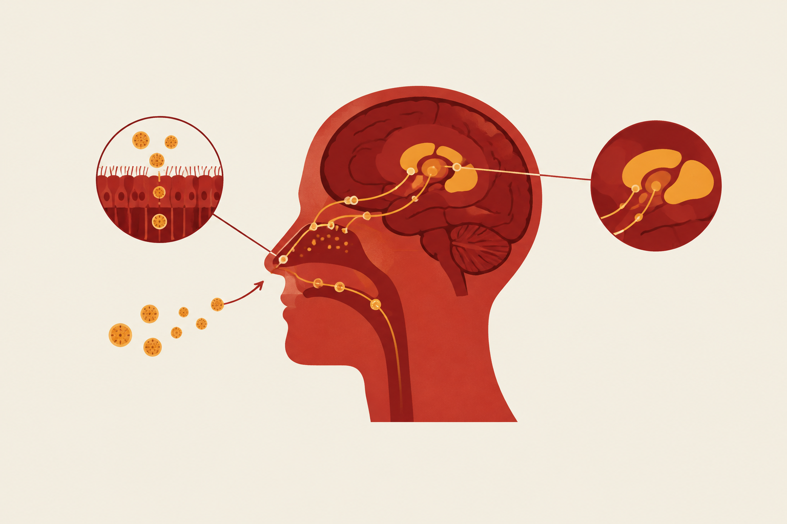
Nanoparticle nose-to-brain delivery for neurological indications uses the olfactory and trigeminal pathways to bypass the blood–brain barrier. Lipid nanoparticles can protect peptide cargo and improve its passage across nasal mucosa, but their destination depends on deposition, particle size, and surface chemistry. For brain-targeted GLP-1 therapeutics, rational design therefore starts with the anatomy linking nasal entry to reward circuits and dementia-relevant structures.
Why the blood–brain barrier makes peripheral injection an inadequate delivery strategy for brain-targeted GLP-1 therapeutics
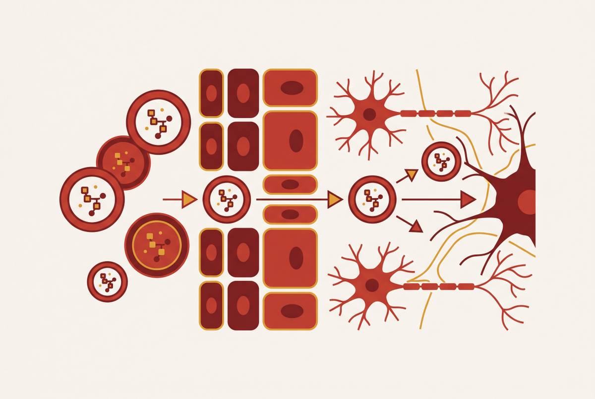
Brain microvascular endothelial cells form continuous tight junctions, supported by pericytes and astrocytic end-feet. Together, they maintain a selectively permeable interface that preserves neuronal homeostasis. This structure severely restricts entry of hydrophilic compounds, peptides, proteins, and antibody-based drugs from the bloodstream. Passive diffusion favors a small subset of hydrophobic, low-molecular-weight molecules, and efflux pumps can return even these to systemic circulation. High plasma exposure does not guarantee brain exposure.
Receptor-mediated transcytosis provides passage for large molecules such as insulin and leptin, but therapeutic GLP-1 formulations do not reliably exploit it at scale. Injection therefore avoids gastrointestinal degradation without solving the central delivery constraint. When a drug is taken by mouth, it faces gastrointestinal degradation and first-pass hepatic metabolism before it even reaches the brain. For addiction, Alzheimer’s disease, and Parkinson’s disease, the drug needs to reach targets inside this protected compartment. When peripheral dosing cannot produce sufficient exposure there, the route is mismatched to the therapeutic objective.
The nasal cavity as a direct anatomical gateway to the CNS
The olfactory epithelium contains bipolar olfactory neurons, sustentacular supporting cells, Bowman’s glands, and basal cells, with a lamina propria underneath. Axonal processes from the neurons form bundles called the filia olfactoria. These pass through the cribriform plate, the perforated structure between the nasal cavity and cranial space, and terminate in the olfactory bulb. The bulb is the brain-side entry point for this anatomical pathway.
The olfactory region occupies a limited part of the nasal cavity, so nasal administration alone does not establish effective olfactory deposition. The trigeminal pathway gives drug another way through, along nerve branches that cover a larger area of mucosa. On a third route, drug is absorbed through nasal blood vessels into systemic circulation and still has to cross the BBB. Intranasal administration avoids gastrointestinal and first-pass hepatic metabolism, but only its direct neural pathways resolve the specific barrier that motivates brain targeting.
The four mechanisms of olfactory transport from epithelial uptake through the cribriform plate to the olfactory bulb
Olfactory transport includes extraneuronal, intraneuronal, transcellular, and paracellular passage. These are distinct mechanisms. In the extraneuronal route, drug moves along the outside of olfactory axons without entering the neurons. In the intraneuronal route, neuronal endocytosis brings material into the cell, followed by transport within the axon toward the olfactory bulb. Transcellular transport through sustentacular cells supplies a separate uptake route through the supporting epithelium, while paracellular transport passes between epithelial cells.
The answer depends partly on its properties, because access to a cellular interior differs from passage outside cells. Neuronal transport also runs in both directions, with anterograde movement toward the brain and retrograde movement in the opposite direction. That bidirectionality complicates assumptions about residence time and how far cargo will travel. Arrival at the bulb is therefore a transport stage.
From the bulb, projection pathways provide access toward deeper structures. Their relevance depends on successful entry upstream. A formulation that lands away from the olfactory region may take a different pathway or miss this route altogether. The mechanistic question is consequently not just whether nasal material enters the brain, but how it crosses the epithelium and proceeds beyond the bulb.
The trigeminal pathway as a parallel highway with distinct anatomy and broader mucosal coverage
Trigeminal branches innervate respiratory epithelium as well as portions of the olfactory region. Their broader coverage allows CNS-directed transport from nasal sites outside the small olfactory area. Here, the conduit is the trigeminal nerve. Its central terminal fields lie in the pons and medulla, giving transported material initial access to brainstem structures. This is a different entry map from olfactory bulb projections toward limbic and cortical regions.
Both pathways can operate after a single intranasal dose. MicroCT nanoparticle tracking has visualized migration along trigeminal as well as olfactory nerves, providing particle-tracer evidence for both routes. Their combined contribution can broaden distribution across CNS compartments, but broader distribution does not mean equivalent access to every target. Brainstem entry is relevant to brainstem-connected circuitry, whereas olfactory entry is more directly aligned with limbic-adjacent destinations. The trigeminal route thus provides anatomical redundancy without substituting for olfactory targeting when the intended destination is hippocampal or cortical.
How do lipid nanoparticles get from the nose to the brain past the mucosal barrier?
Before either neural pathway becomes available, a peptide must survive the nasal mucosa. Mucociliary clearance moves mucus toward the nasopharynx, removing deposited material from the uptake site. Proteases and other enzymes can degrade peptide cargo during that interval. Simple drops, sprays, and powders may offer only a short residence time, while peptides themselves have poor epithelial permeability without a carrier. Anatomical access alone is therefore not enough.
Lipid nanoparticles address these obstacles through protection, retention, and permeation. Encapsulation can shield the payload from enzymatic degradation, while lipid-based formulations can improve solubility and epithelial passage. Mucoadhesive surfaces extend contact with the mucosa, increasing the opportunity for uptake before clearance. These properties make LNPs a strong engineering fit for nasal peptide delivery. Even with these properties, lipid formulations can differ in how well they reach a given brain region.
The lipid-based platforms under development include liposomes, nanoemulsions, solid lipid nanoparticles, and nanostructured lipid carriers. Their differing lipid structures affect loading, release kinetics, and surface presentation. Solid lipid nanoparticles and nanostructured lipid carriers can improve solubility and permeation while prolonging drug action and reducing degradation. Particles smaller than 1000 nm have a high surface area and may offer better absorption and stability than conventional formulations. That broad size category describes a useful vehicle class.
Particle size, surface chemistry, and the engineering decisions that determine which brain region an LNP reaches
Aerosolized quantum dots in the ~15 nm range have demonstrated olfactory axonal uptake, connecting very small particle size with the intraneuronal route and olfactory bulb entry. Coated gold–iron oxide nanoparticles below 50 nm have shown migration along both olfactory and trigeminal nerves, with additional paracellular transport. Larger particles may depend more heavily on extraneuronal and paracellular passage. Size changes the available transport options and, through them, the initial CNS destination.
These examples establish mechanisms using particular tracer materials. Surface chemistry supplies another determinant of transport. Lactoferrin-trimethyl chitosan, or Lf-TMC, surface modification has enhanced nanoparticle brain targeting and prolonged residence, with accumulation in the olfactory bulb and hippocampus. Chitosan and similar mucoadhesive polymers also extend epithelial contact before mucociliary removal.
Hippocampal accumulation aligns a formulation with a primary site of early Alzheimer’s neurodegeneration. For reward circuits, efficient olfactory bulb entry provides access toward limbic networks and downstream structures, but entry alone does not establish their exposure. Stimuli-responsive surfaces are a new way to further adjust delivery. Design must identify the combination of size, coating, and deposition that favors the route to the intended destination, rather than the formulation that produces the largest undifferentiated brain signal.
GLP-1 receptor distribution in reward circuits and dementia-relevant structures and why the destination matters
GLP-1 receptors are present in the ventral tegmental area, nucleus accumbens, and prefrontal cortex. These regions participate in reward, motivation, and the control of behavior. Receptor activation influences dopaminergic, glutamatergic, and GABAergic neurotransmission within these systems. Preclinical GLP-1 receptor agonist studies have attenuated intake and relapse-like behavior involving alcohol, nicotine, and cocaine.
For dementia, the hippocampus and neocortex are key destinations because they are vulnerable in Alzheimer’s disease. Their GLP-1 receptor expression gives a drug delivered to these regions a plausible biological target. Insulin resistance, metabolic failure, and neuroinflammation also link how metabolism works with the pathology of Alzheimer’s and Parkinson’s disease. Observational associations between GLP-1 receptor agonist use and lower dementia incidence strengthen the rationale for investigation. They do not establish that targeting these regions will produce clinical benefit.
Olfactory bulb projections connect toward the amygdala, entorhinal cortex, and hippocampus, with downstream influence on the ventral tegmental area and nucleus accumbens through multisynaptic pathways. Trigeminal brainstem entry instead aligns with brainstem-connected circuitry. In rodents, selected intranasal agonists have shown preferential access to hippocampus and neocortex, although hypothalamic uptake is higher overall. Thus, receptor location, anatomical connection, and measured drug exposure are related questions, but none substitutes for the others.
What preclinical intranasal GLP-1 data reveal about the gap between peripheral injection and direct brain targeting
Intranasal studies in CD-1 mice and APP/PS1 Alzheimer’s disease models found widespread brain distribution of dulaglutide, exenatide, and the dual agonist DA4-JC. Dulaglutide and DA4-JC stood out for access to hippocampus and neocortex shortly after administration, while overall uptake favored the hypothalamus. This rapid regional distribution is consistent with direct olfactory and paracellular transport. On its own, it cannot show how much each pathway contributes. Nor does a peptide distribution study establish that an LNP formulation will reproduce the same profile.
Liraglutide’s ELAD phase 2 trial used subcutaneous delivery and reported reduced gray matter volume loss, including in the temporal lobe and whole cortex, with attenuated FDG-PET decline over 12 months. Yet it did not meet its primary cerebral glucose metabolism endpoint. The EVOKE and EVOKE+ trials tested oral semaglutide in thousands of participants with mild cognitive impairment or mild Alzheimer’s disease. They showed no significant slowing of disease progression even though biomarkers improved, and safety and tolerability were in line with prior studies.
Parkinson’s trials have produced motor benefits with exenatide and lixisenatide in some studies, while NLY01, a pegylated exendin-4, did not meet its primary endpoint. Limited brain penetration and gastrointestinal side effects that restrict dose escalation create a plausible exposure bottleneck, even when plasma levels are high. However, the controlled trial outcomes do not prove that inadequate delivery caused the failures. Preclinical nose-to-brain pharmacokinetics support testing that explanation.
Why formulation design guided by pathway anatomy produces better-targeted LNPs than iterative empirical testing
An endpoint brain concentration cannot distinguish clearance before uptake from unsuitable pathway engagement or inadequate regional targeting. Each failure suggests a different formulation problem. Design guided by pathways starts with the region the drug needs to reach, such as hippocampus for Alzheimer’s disease or reward circuitry for addiction. It then matches that destination to olfactory projections or trigeminal brainstem entry, and selects particle dimensions and surface modifications to support the relevant transport mechanism. Iterative testing remains necessary, but anatomy gives each iteration an interpretable hypothesis.
Human translation is the critical unresolved step. Rodent and human olfactory anatomy differ in surface area and airflow, so successful rodent uptake cannot simply be transferred to human dosing. In vitro and ex vivo nasal models that better reproduce human deposition, patient-specific delivery platforms, and smart nanoparticle surfaces provide ways to investigate that gap. Their role is to connect deposition and retention with pathway engagement and regional exposure. A better-targeted formulation must ultimately demonstrate that connection.
The platform insight extends beyond GLP-1 peptides. The same anatomical knowledge can inform nasal delivery of other peptides, gene editing cargo, and regenerative medicine payloads, although their formulations still require separate evaluation. For neurological indications where peripheral injection cannot achieve sufficient CNS exposure, that transferable mechanistic foundation could make a treatment feasible where systemic delivery alone cannot.
Sources
- Nanocarrier-based intranasal drug delivery for enhanced neurological disorders treatment
- Frontiers
- Advancements in Nanocarrier Systems for Nose-to-Brain Drug Delivery
- Pharmacokinetics and Pharmacodynamics of Intranasal Solid Lipid Nanoparticles and Nanostructured Lipid Carriers for Nose-to-Brain Delivery
- Formulation considerations in enhancing olfactory mucosal deposition for nose-to-brain drug delivery
- Lipid-based nanoparticles via nose-to-brain delivery: a mini review
- Intranasal delivery of therapeutics to degenerating brain regions: From pathways to nanocarriers - ScienceDirect
- Nose-to-brain drug delivery: from bench to bedside